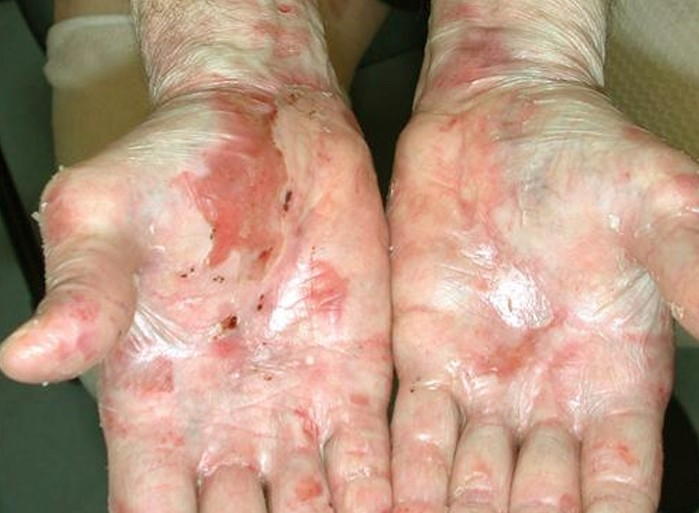
Epidermolysis bullosa is a blistering skin disease which is usually first noticed during early childhood. In epidermolysis bullosa, blisters form on the skin following minor skin trauma, such as bumping into objects, sitting on hard surfaces and walking. This is because the skin of sufferers of epidermolysis bullosa is highly fragile. It is a rare genetic condition, and there is a positive family history of the disease in many patients who are diagnosed with the disease.
There are three main types of epidermolysis bullosa which are classified according to the level of the skin which is affected. The deeper the level of the skin affected, the more severe the disorder.
The three subtypes are:
- Epidermolysis bullosa simplex: this form of epidermolysis bullosa affects predominantly the hands and feet. This type of the condition is caused by genetic defects in keratin, a protein which is an important structural component of the skin. This form of the condition affects the epidermis, which is the top layer of the skin. The blisters which form tend to heal quite well, and generally do not scar.
- Junctional epidermolysis bullosa: This is one of the rarer types of epidermolysis bullosa, and as the name suggests, it affects the layer of the skin between the epidermis and the dermis. This layer is called the basement membrane. This form of the disease is the result of abnormalities in collagen (a type of connective tissue) and laminin (a protein found in the basement membrane which helps to hold cells and tissues together).
- Dystrophic epidermolysis bullosa: this type of the disease affects the dermis, the deepest skin layer. There are also several subtypes of dystrophic epidermolysis bullosa. The dystrophic epidermolysis bullosa subtypes often produce scarring after the blisters have healed. The nails, scalp and teeth are also affected, as are many internal organs. Scarring also affects the internal mucous membranes, which can lead to strictures (narrowing) of internal passages, such as the oesophagus and urethra. Dystrophic epidermolysis bullosa tends to be due to genetic mutations affecting the collagen in the dermis.
The blistering is severe and very prone to infection, as the wounds heal poorly. The long-term skin inflammation caused by dystrophic epidermolysis bullosa is problematic, as it can damage the DNA within the skin cells. This greatly increases the risk of developing squamous cell carcinoma. The life expectancy of sufferers of dystrophic epidermolysis bullosa is approximately 30 years; with death usually resulting from complications of the disorder itself or due to squamous cell carcinoma. The skin of children with this condition is very fragile, and for this reason, children with the disorder are sometimes called “Butterfly Children)
There are several subtypes of epidermolysis bullosa, which have varying degrees of severity:
Mild forms: The most common subtype is called the Weber-Cockane subtype, and this is the mildest form of the disease. Blisters are mainly found on the palms of the hands and the sole of the foot, and in most instances were caused by some form of trauma to the area. This subtype is sometimes also associated with hyperhidrosis, which is a condition which causes excessive sweating.
Severe forms: In the more severe subtypes of epidermolysis bullosa simplex (Koebner subtype and Dowling-Maera subtype), the blisters are more widespread and the onset of blistering is first noticed a short period of time after birth. Particularly in the Dowling-Maera subtype, the blisters may affect the mucous membrane lining the mouth. In the Koebner subtype, patches of the skin, particularly of the palms and soles, become thickened and the pigmentation of the skin appears darker.
There are 2 main subtypes of the condition:
Herlitz subtype: This is the most severe form of junctional epidermolysis bullosa. This subtype is often fatal and children affected by the condition generally do not survive beyond infancy, because they are very prone to infections which can enter the bloodstream. Additionally, there are also respiratory problems because the condition affects both the skin and internal organs. On the skin, there are obvious erosions surrounding the mouth, nose, eyes and anus. It also affects the lining of the gastrointestinal tract, from the mouth to the rectum, the lining of the respiratory system, the eyes and the lining of the genitourinary system.
Non-Herlitz subtypes: These subtypes cause deeper blisters and erosions than what are seen in epidermolysis bullosa simplex. The blisters are quite widespread across the body. The non-Herlitz forms may also affect the nails, the scalp and the teeth. There may be some involvement of internal organs, particularly of the gastrointestinal tract, with the non-Herlitz subtypes.
Symptoms and Signs
The signs and symptoms of the disease vary depending on the subtype.
Some possible signs and symptoms of the condition include:
- Development of fluid-filled blisters on the skin after some form of skin trauma. The blisters are typically found on the palms and soles
- Thickening of the skin on the palms and soles
- Scarring of the skin
- Changes to the fingernails and toenails
- Blistering and scarring of the scalp; this may also be accompanied by hair loss
- Tooth decay and other dental problems
- Difficulty swallowing
- Constipation
- Growth and developmental delay
Complications
There are multiple complications which are associated with epidermolysis bullosa. The potential complications vary with the subtype and the severity of the disorder. The most common complication of epidermolysis bullosa is infection. Having intact, unbroken skin is one of our most important defences against infection, because the skin has many defences that can stop bacteria and viruses from entering.
The blisters and erosions break the skin and it is very easy for bacteria and viruses to enter the skin and cause infection. Infections of the wound can then also spread to the bloodstream and result in sepsis; which is a very serious and potentially fatal complication if left untreated. The skin is also an important organ because it helps to prevent loss of fluid. Patients with epidermolysis bullosa are prone to dehydration, because the breaks in the skin allow fluid to escape easily. As mentioned above, chronic inflammation in the skin can induce DNA mutations in skin cells, which greatly increases the risk of developing squamous cell carcinoma. Particularly with subtypes affecting the deeper skin layers, contractures can form, which can lead to impaired joint function and deformity.
Complications can also arise from internal organ involvement. Due to involvement of the gastrointestinal tract, there are often difficulties with feeding and nutrition. If the lining of the mouth and oesophagus are affected, eating and swallowing is painful, and it is difficult for the sufferer to eat and drink sufficiently. Additionally, scarring of the oesophagus can result in narrowing, which also makes it difficult for food to pass down into the stomach. Due to reduced intake of food and drink, children can become malnourished, which can lead to problems with growth and development.
Good nutrition is important for skin healing, so this can lead to an added problem of slow wound healing. If intake of fibre is inadequate, this can contribute to constipation, which is often already an issue due to blistering and narrowing because of scarring. In some forms of the disease, the eyes can be affected, which can lead to visual problems and potentially blindness in severe circumstances.
Treatment
Treatment of epidermolysis bullosa is aimed at treating symptoms and complications. There are no medications or treatments which can treat the underlying causes.
Some possible treatments for the disorder include:
- Antibiotics: antibiotics are necessary to treat infected wounds and to help prevent the spread of the infection into the bloodstream
- Wound care: dressing and covering wounds is also important to promote healing and to prevent infection
- Oral care: good oral hygiene and regular dental check-ups are important for patients with involvement of the teeth and oral cavity
- Nutritional/dietary supplements
- Corticosteroids and phenytoin are medications which can be used to ease some of the symptoms arising from the oesophagus. However, there are many side effects of corticosteroids and they should not be used as a long-term treatment
- Surgery may be required to dilate the oesophagus and relieve strictures. This is achieved by dilating the oesophagus with a balloon. Surgery may also be required to correct contractures and remove skin cancers.


