
Tinea capitis, otherwise known as scalp ringworm, is a fungal infection of the scalp. It is more prevalent among young children than adults. The fungi causing the infection can be caught from animals such as dogs, cats, guinea pigs and cattle. Some people are carriers of tinea capitis. These people do not have any symptoms of the infection, but the fungus is still present on the scalp.

Causative Organisms
Tinea capitis is an infection caused by a specific group of fungi called the dermatophytes. Dermatophytes need keratin, a protein which is a key component of our hair, skin and nails, to survive. Hence, these fungi typically cause superficial infections of these sites. Two classes of dermatophytes that are implicated in tinea capitis infections are Microsporum (M.) and Trichophyton (T.). M. canis is the most commonly implicated organism in tinea capitis transmitted by animals. Cats and dogs are common sources for this organism, particularly kittens and puppies. Other organisms implicated in tinea capitis infections include T. verrucosum, which is found in cows, and M. nanum, which is found in pigs.
Risk Factors
Some of the risk factors for developing a tinea capitis infection include:
- Age: tinea capitis is most common in young children. The condition becomes less common after puberty. This is because of the protective role of sebum. Sebum is the greasy substance found on skin and hair that is secreted by the hair follicles. The secretion of sebum greatly increases during puberty. The acidity of the sebum is protective against tinea capitis because it inhibits the growth of the fungi which cause the infection. Younger children also tend to have poorer hygiene and spend time with pets and other animals which carry the causative organisms.
- Poor personal hygiene
- Overcrowding
- Low socioeconomic status
- Ethnicity: tinea capitis is more common in indigenous people
Types of Tinea Capitis
There are 3 main types of tinea capitis infections which have been described; ectothrix, endothrix and favus. Ectothrix infections are typically what occur when the causative organism is of the Microsporum family. The outside of the hair shaft is affected by the fungus (ecto- = outside). Endothrix infections are caused by Trichophyton fungi, and in these infections the fungi infect the inside of the hair shaft (endo- = inside). Favus is a less common type of infection in Australia. It is more common in Africa and the Middle East. It is typically caused by T. schoenleinii. It also affects the inside of the hair shaft, where there are airspaces between the branches of the fungus (called hyphae), giving the inside of the hair what is sometimes described as a honeycomb appearance. Favus is a severe form of tinea capitis that presents with matted hair and yellow, crusty lesions.
Appearance
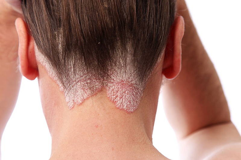
Tinea capitis can be divided into inflammatory and non-inflammatory types. Non-inflammatory tinea capitis gives the scalp a scaly appearance (non-inflammatory tinea capitis can also be called seborrhoeic due to the scale). Non-inflammatory tinea capitis is caused by organisms including M. ferrugineum and M. audouinii. These organisms grow on the outside of the hair and cause the hairs to turn grey. These hairs are brittle and fall off easily, resulting in black spots where the hairs have broken. Surrounding hairs are also fragile and can fall out easily. The areas of hair loss (alopecia) tend to be well-defined. In the overwhelming majority of cases, the alopecia is temporary and the hair grows back. The patches of alopecia tend to be most common on the back of the head (the region is known as the occiput).
Other dermatophytes cause an inflammatory type of tinea capitis (M. canis, T. verrucosum). Kerions are typical lesions seen in inflammatory types of tinea capitis. Kerions are lesions that are the result of the inflammatory response that may look like an abscess. They are red and hot, and feel boggy to the touch. There may be a discharge coming from the kerion. The kerion may develop a crusted appearance in later stages. Scarring alopecia may develop. In inflammatory forms of the condition, the scalp is very tender and quite itchy. Enlargement of the lymph nodes in the neck is also a common finding in inflammatory forms; this is the result of the immune system attempting to clear the infection.
Examination
In suspected cases of tinea capitis, a Wood Lamp is used to examine the scalp and hairs. The Wood Lamp emits UV light that is not visible to the human eye (black light). The Wood lamp is used to examine the skin and scalp. The different types of tinea capitis appear differently when viewed with the Wood Lamp:
- Ectothrix: ectothrix infections glow when examined with the Wood Lamp. If canis is the organism causing the infection, the glow will be a green colour
- Endothrix: endothrix infections are not fluorescent under examination with a Wood lamp
- Favus: schoenleinii infections glow a green or blue to white colour
Treatment
Treatment of tinea capitis needs to be given orally, as topical treatments often cannot access fungi within the hair follicles. There are several types of antifungal medications that can be used in the treatment of tinea capitis. These include:
- Griseofulvin: this is a medication that can be used in patients 4 years of age and older. It works by inhibiting mitoses in fungi; this is achieved by affecting the function of microtubules. It needs to be administered for 6-8 weeks; hence there are issues with patients adhering to the treatment regime. It can cause headaches, light sensitivity, nausea and vomiting.
- Terbinafine: terbinafine is an antifungal that works by inhibiting enzymes needed to make the cell membrane of fungi, resulting in lysis of the fungal cells. It can eradicate infections with Trichophyton within 4 weeks, and can eradicate Microsporum infections within 8 weeks. Side effects include nausea, vomiting, diarrhoea, headaches, fatigue and taste disturbances.
- Fluconazole: fluconazole is an antifungal medication that works by inhibiting fungal enzymes needed in the process of synthesising the cell membrane. Common side effects include nausea, vomiting, diarrhoea, headaches and rashes. It can be given as a tablet or as a syrup. Treatment with this medication can be as short as 20 days in duration.
- Itraconazole: as with fluconazole, itraconazole inhibits the formation of the fungal cell membrane by blocking enzymes required in cell membrane synthesis. Treatment takes 2-4 weeks to eradicate the infection. Potential side effects include diarrhoea and gastrointestinal upsets, as well as changes to liver enzymes.
If you have any questions or concerns about scalp tinea or scale, contact your local doctor, who will arrange for you to see a dermatologist. Contact us today.