
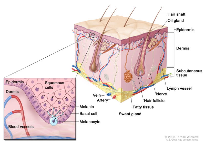

The skin is the body’s largest organ. It has two main layers which serve a number of important functions. These layers are called the epidermis (the outer layer) and the dermis (the middle layer). The subcutaneous tissue lies below the skin.
Functions of the Skin
The skin serves many important functions, which include:
- Protection: the skin acts as a barrier to prevent bacteria, viruses and fungi from entering the body to cause an infection. People who have suffered from severe burns are at a greatly increased risk of infection because the integrity of this vital barrier is compromised. It helps shield the delicate tissues underneath from mechanical and other injuries.
- Excretion: the skin is a site through which we can excrete urea and other wastes via the sweat (the skin is not as important as the kidneys in terms of excretion)
- The skin is an important part of the sensory system. It contains important receptors which are specialised to detect touch, pressure, heat, cold, vibration and pain
- The skin is also very important for the control of our body temperature. It is the site of sweating, which helps to remove heat from the body. The skin also has a lot of blood vessels. These blood vessels constrict (become narrower) when the body temperature drops to help conserve heat, and dilate (become wider and increase blood flow) when body temperature increases to help release more heat via the skin
- The skin helps prevent us losing fluid as it is quite waterproof
- It guards against excessive exposure to the ultraviolet rays of the sun by producing a protective pigmentation called melanin
- The skin is also a site of storage of fats
- The skin is involved in the synthesis of vitamin D, an essential vitamin which is obtained from sunlight
The Epidermis
The epidermis is the outer layer of the skin. It itself has a number of different layers. It is a waterproof protective layer made up of many different cells. From the bottom upwards, the layers of the epidermis are:
- Stratum Basale: this is the deepest layer of the epidermis and is only one layer of cells thick; although it may be 2-3 layers thick in areas of skin without hair. It is made up of cells called keratinocytes, which are like the stem cells of the skin. These cells divide into 2 and ascend superficially to the next layer of the skin, called the stratum spinosum. It also has melanocytes, which are pigment cells that produce melanin, which gives the skin its colour. Touch receptors, called Merkel’s cells are responsible for detecting the sensation of light touch and two-point discrimination. Another important cell type found in the stratum basale is the Langerhan’s cells, which are a part of the immune system. They are antigen-presenting cells, which mean that they detect foreign bacteria, viruses and fungi and present these to cells of the immune system to mount an immune response
- Stratum Spinosum: this is the layer where the cells start to become filled with keratin. This layer is sometimes called the prickle cell layer because the cells look ‘spiky’ due to connections between adjacent cells
- Stratum granulosum: this is a thin layer. The cells in this layer are filled with granules that are needed to bind filaments of keratin together within the cell
- Stratum lucidum: this layer is only present in areas of thick skin, namely, the palms of the hands and the soles of the feet. It is a layer that is 3-5 cells thick, containing flat keratinocytes.
- Stratum Corneum: this is the outer layer consisting of keratinised dead squamous skin cells. It acts as a barrier to prevent the entry of potentially dangerous bacteria, fungi and viruses, as well as to water-proof the skin. It takes about 2 weeks for the skin cells to migrate from the stratum basale to the stratum corneum. Eventually, skin cells are shed from the stratum corneum into the environment. This is the thickest layer of the epidermis, and can be up to 30 cells thick.
The epidermis is avascular – it has no blood vessels directly in the layer. Veins and arteries are found just deep to the epidermis. Nutrients and oxygen diffuse into this layer from the bloodstream.
Dermis
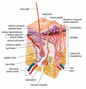

The top layer of the dermis is called the papillary dermis, so named for the finger-like projections (papillae) which project upwards to the epidermis. It is very vascular, containing lots of blood vessels which supply the dermis and indirectly, the epidermis. It also contains a special type of receptor, called Meissner’s corpuscles. Meissner’s corpuscles are specialised nerve endings which detect touch and vibration. Within the papillary dermis, the main type of connective tissue is called loose connective tissue. The loose connective tissue here is strong enough to hold the dermis, epidermis and blood vessels together. Since it is not very rigid, it is also flexible and helps to cushion the skin; providing a healthy medium of both strength and elasticity.
The deeper layer of the dermis is called the reticular dermis. It is much thicker than the papillary layer and contains a number of important structures. The main type of connective tissue in the dermis is dense, irregular connective tissue which is much stronger than the loose connective tissue in the papillary dermis. The reticular dermis provides most of the overall strength of the skin. Also located in the reticular dermis are the sweat glands and hair follicles. The sweat glands have ducts which open up onto the surface of the skin. In the reticular dermis, there is another type of touch receptor, called Pacinian Corpuscles. These corpuscles are involved in the detection of deep touch and vibrations. The reticular dermis also contains larger blood vessels than those found in the papillary dermis.
Subcutaneous Tissue (Hypodermis)
The hypodermis is under the dermis. It contains a lot of adipose (fatty) tissue, as well as macrophages (cells of the immune system) and collagen. It also contains blood vessels and nerves. Due to the number of blood vessels, the hypodermis is the layer of the skin that insulin and other therapeutic drugs are injected into to increase absorption. The main function of the hypodermis is to store fat – up to half of the body’s fat is stored in the hypodermis. The fat in this layer is important to provide insulation to help keep the body warm and it is also involved in shock absorption and energy storage.
If you have any questions on this topic or others you may Contact us today.